Cannabis under the scanning electron microscope

          Amazing photo of cannabis under the scanning electron microscope was a dome shape like
football ( pink colour) of the female cannabis (Cannabis Thai Stick) from leaves and petiole. The
cannabis plant has had a complicated relationship with humans through the ages and each image was
present highlighting unique and bizarre features. This photographic revealed the microscopic features
of the cannabis plant as never seen before.

          A scanning electron microscope image of a nice elongated trichome structure on the top of a
cannabis leaves as following show cell structures act as thorns on the leaves top.

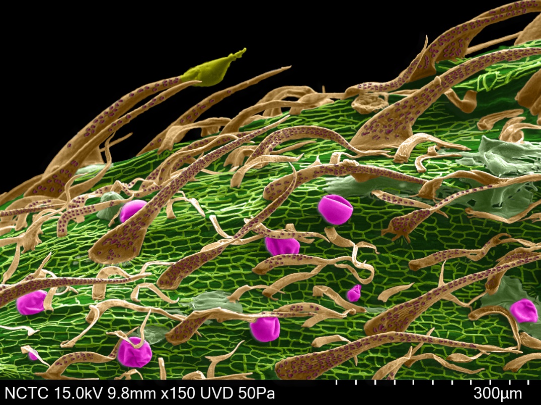
Figure 1. SEM image of Cannabis petiole at 150X.

          The oval bract structure houses the stigma that responsible for producing seeds when the flower has been pollinated. This bract structure was the highest concentration of cannabinoid compounds on the plant that located the highest concentration of THC (Tetrahydrocannabinol).

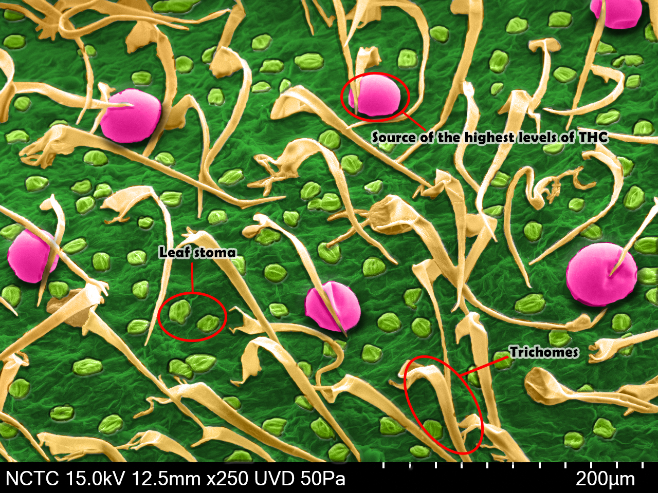
Figure 2. SEM image of Cannabis leaves at 250X.

          A sample at the bottom of cannabis leaves found two types of defensive cell structures, first: the tall needle like trichomes (physical defence) and second: the football like glandular trichomes (chemical defence) were the sources of the highest levels of THC. The high density of trichomes on modern cannabis leaves lead to good sources of THC levels.

Figure 3. SEM image of bottom of cannabis leaves at 200X.

          The difference SEM image colours shown various components from the central rib at the bottom of a leaves towards the edge of the blade. This light green colour on SEM image of the cell structure allowed the carbon dioxide to enter inside the leaves. The leaves stoma made of two cells called the guard cells that opened and closed depending on the time of day.

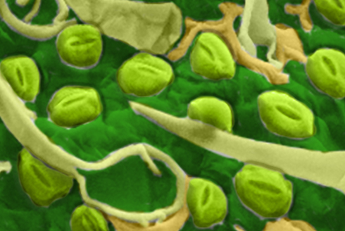
Figure 4. SEM image of Cannabis leaves stoma.

          As the highest levels of THC have studied in plant defence insect and herbivore predators then a human use this chemical in medical interest and develop the plant to increase the THC levels. THC is shorthand for Tetrahydrocannabinol also known as ((6aR,10aR)-delta-9-tetrahydrocannabinol), it is the psychoactive chemical in the cannabis plant. The closely image on the surface shown stoma structures scattered around the leaves surface that allow leaves breathing.

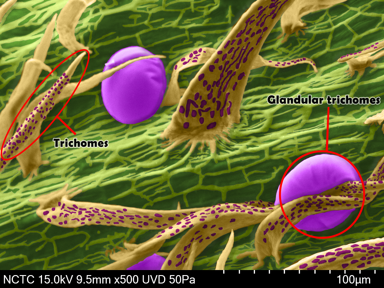
Figure 5. SEM image of Cannabis petiole at 500X.

          The studied of THC concentration with scanning electron microscope would be helpful for provided an information for identification of THC levels before analysis with another method that may be difficult for preparation or take a lot of time for analysis.

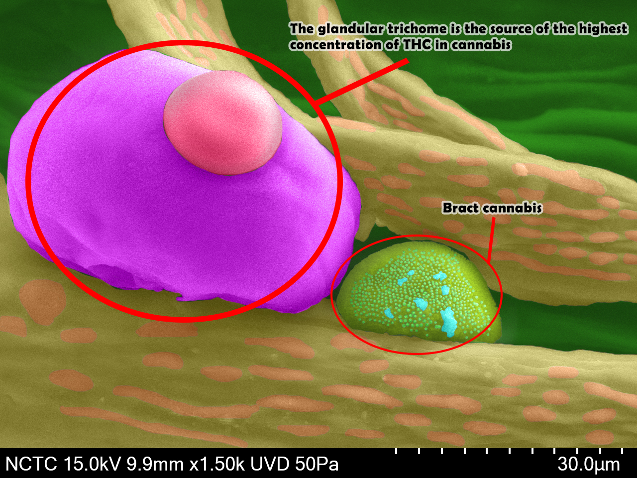
Figure 6. SEM image of Cannabis vein leaves* at 1,500X

*Remark:
An individual glandular trichome (purple and pink colour), the glandular trichome is the source of the highest concentration of THC in cannabis most often found on the bottom of the leaf and in the plant bud.)

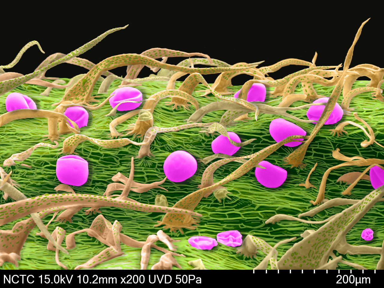
Figure 7. SEM image of Cannabis midrib leaves at 200X

References

[1] https://www.businessinsider.com/marijuana-cannabis-scanning-electron-microscope-2018-9
[2] https://www.snopes.com/fact-check/marijuana-electron-microscope/
[3] https://www.mic.com/articles/168526/marijuana-under-a-microscope-these-ultra-close-up-images-might-just-blow-your-mind0
[4] https://www.researchgate.net/publication/248568268_Structural_investigation_of_the_glandular_trichomes_of_endemic_Salvia_smyrnea_L
[5] https://fineartamerica.com/featured/sage-leaf-trichomes-sem-peter-bond-em-centre-university-of-plymouth.html
[6] http://www.scielo.br/scielo.php?script=sci_arttext&pid=S0100-84042008000300010

Prathompoom Newyawong
Laboratory Officer, Microscopy Laboratory,
NSTDA Characterization and Testing Service Center (NCTC)
E-mail: prathompoom.newyawong@nstda.or.th